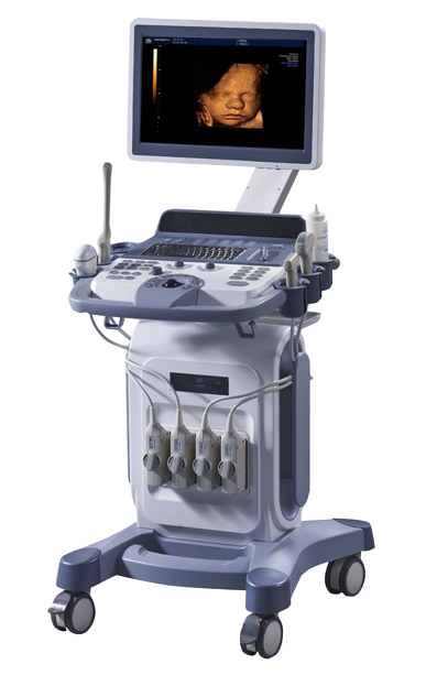
Color Doppler
A Doppler ultrasound, also called a Color Doppler test is a non-invasive test that can be used to estimate your blood flow through blood vessels. It helps doctors evaluate blood flow through major arteries and veins, such as those of the arms, legs, and neck. It can show blocked or reduced flow of blood through narrow areas in the major arteries of the neck that could cause a stroke. It also can reveal blood clots in leg veins (deep vein thrombosis) that could break loose and block blood flow to the lungs (pulmonary embolism). During pregnancy, Doppler ultrasound may be used to look at blood flow in an unborn baby (foetus) to check the health of the foetus.
A Doppler ultrasound can estimate how fast blood flows by measuring the rate of change in its pitch (frequency). During a Doppler ultrasound, a doctor trained in ultrasound imaging (sonographer) presses a small hand-held device (transducer), about the size of a bar of soap, against your skin over the area of your body being examined, moving from one area to another as necessary.
This test may be done as an alternative to more-invasive procedures, such as angiography, which involves injecting dye into the blood vessels so that they show up clearly on X-ray images.
A Doppler ultrasound test may also help your doctor check for injuries to your arteries or to monitor certain treatments to your veins and arteries.
A Doppler ultrasound may help diagnose many conditions, including:
- Blood clots
- Poorly functioning valves in your leg veins, which can cause blood or other fluids to pool in your legs (venous insufficiency)
- Heart valve defects and congenital heart disease
- A blocked artery (arterial occlusion)
- Decreased blood circulation into your legs (peripheral artery disease)
- Bulging arteries (aneurysms)
- Narrowing of an artery, such as in your neck (carotid artery stenosis)
