3D-4D Sonography
Doctors use ultrasound in women, men, and children to gain advanced insights into the inner workings of the body. In fact, after x-ray exams, ultrasound is the most utilized form of diagnostic imaging available today.
Ultrasound can help diagnose a wide variety of conditions in men, ranging from heart disease to abnormalities in the prostate gland or testicles. With children, doctors commonly use ultrasound to detect a variety of illnesses and disorders. A physician may use ultrasound to examine a child's gastrointestinal tract for signs of appendicitis or a baby's bone structure for alignment problems like congenital hip dislocation or spina bifida. An ultrasound exam of the head can detect hydrocephaly (water on the brain), intracranial hemorrahage (bleeding in the skull), and other conditions of the head.
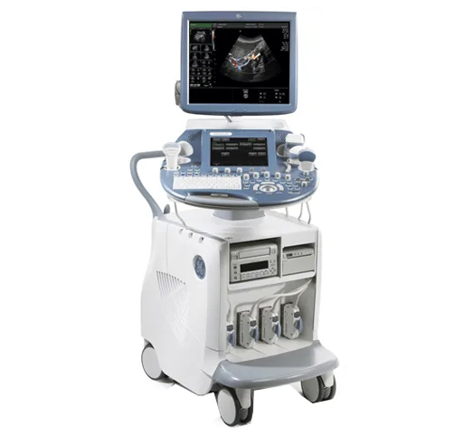
Despite today's sophisticated, high-tech systems, ultrasound remains a science built upon the simple sound wave. By beaming high-frequency sound waves into the body, physicians translate the echoes that bounce off body tissues and organs into colorful, visual images that provide valuable medical information. Heart disease, stroke, abnormalities in the abdomen or reproductive system, gallstones, liver damage, and kidney dysfunction all exhibit telltale signs that ultrasound can help to detect.
Safe, affordable, and non-invasive, ultrasound is also portable. Ultrasound helps doctors make a diagnosis and determine the best and most effective means possible to achieve health.
Sonography
Parents often want 3D and 4D ultrasounds. They let you see your baby’s face for the first time. Some doctors like 3D and 4D ultrasounds because they can show certain birth defects, such as cleft palate, that might not show up on a standard ultrasound.
On the day of the test
It is very important to arrive with a full bladder. This allows the technologists and radiologist to view the bladder while it is full and after it has been emptied. Your ultrasound test is performed by registered, expert radiologists. You may be asked to change into a hospital gown.
How the Test Is Done
For an abdominal ultrasound, you’ll lie down and a technician will put a certain gel on your belly. This helps carry the sound waves. Then the radiologist will hold a probe against your belly and move it around to get an image.
What to Know About Test Results
Afterward, you may get photos/CD or a copy of a 4D movie to take home. our doctor will inform you if anything seems unusual.